Treating Tear Trough Deformities
The tear trough is an important area when it comes to aging, as many concerns can arise in this area that needs treatment using ophthalmology medications. For instance, the hollowing of the under-eye area due to volume and fat loss can lead to the appearance of perennial exhaustion in a patient’s face, even if they are well-rested. Because of this, optimal treatment of this area can greatly satisfy a patient, as it instantly gives them a refreshed, youthful look. However, treatment of this area requires intricate knowledge of the anatomy and dermal filler rheology in order to produce satisfactory results .
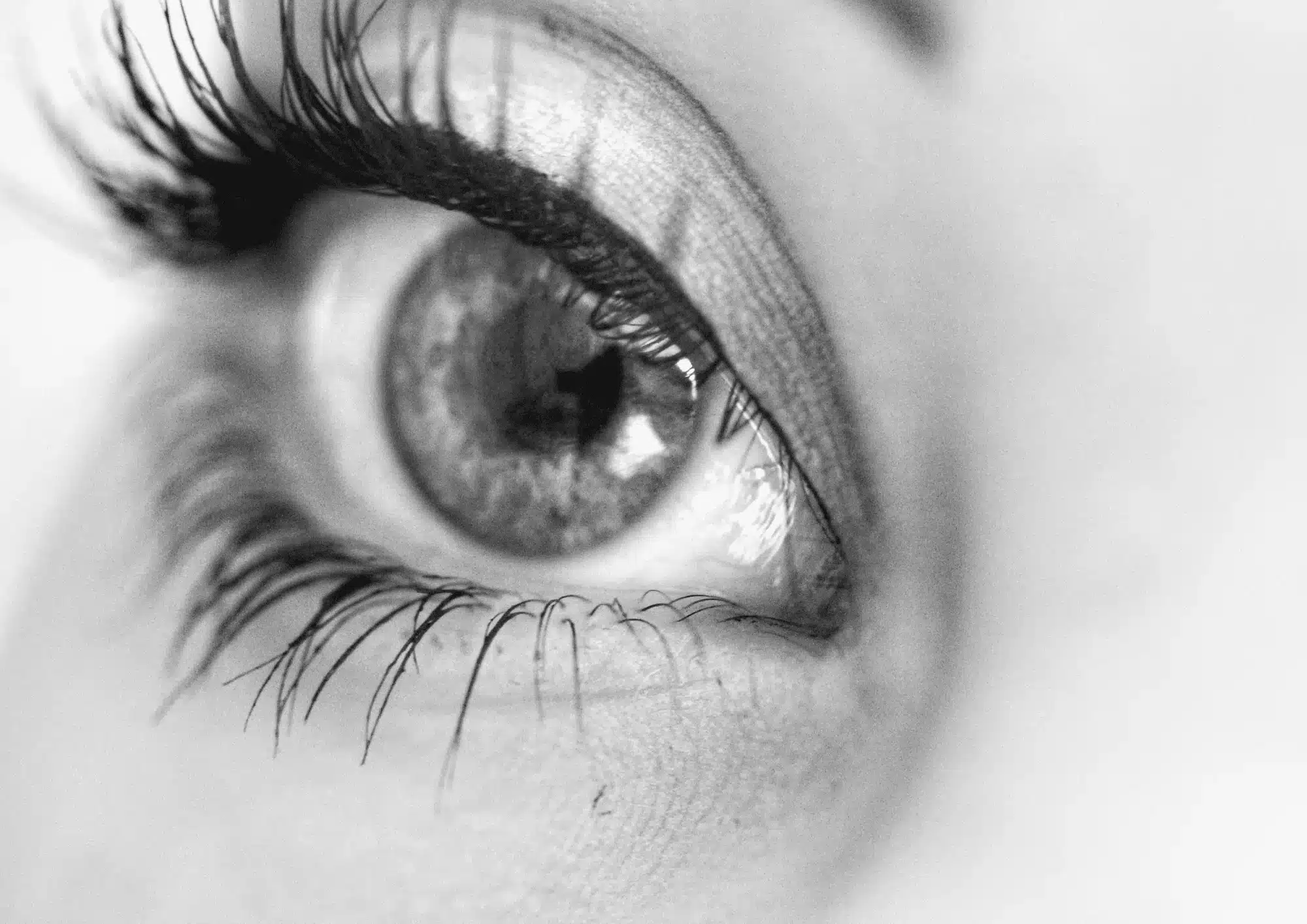
First termed by Flowers in 1993, what is known as the tear trough deformity is a periorbital hollow that extends obliquely from the medial canthus to the mid-pupillary line. Skin depressions lateral to this feature are termed a palpebromalar groove, nasojugal groove, or lid-cheek junction.
There are various classifications of tear trough deformities and its aetiology. For instance, the Tear Trough Rating Scale (TTRS) is an assessment scale that quantitatively assesses tear trough deformities through the measurement of volume of prolapsed fat, skin rhytidosis, depth of the tear trough, and hyperpigmentation. Having a quantifiable measurement of severity helps patients gain a better understanding of the results and the expected outcomes of treatment. It also helps practitioners choose suitable modalities for treatment. For example, with a patient who has a higher degree of pigmentation than tear trough depth, it may be better for the patient to pursue additional treatments, such as skin peels and topical tyrosinase inhibitors, to address the issue(s) and achieve the desired effects.
Tear trough case study
A case study conducted by Dr. Tayyab Bhatti discovered the most optimal approach to treating tear trough deformities. A patient complaining of dark circles under her eyes was seeking treatment for her tear trough region. She was of Middle Eastern descent and was 29 years of age. She was in good health, with no prior medical or surgical history and no known allergies. Previous treatments for her under-eye circles included the use of over-the-counter eye creams, but these were ineffective. A facial assessment showed a degree of hyperpigmentation of the skin under the eye and the presence of a mild to moderate, clearly defined tear trough deformity with the presence of a palpebromalar groove, which served to worsen the appearance of her dark circles. The tear trough deformity was made more pronounced when the patient gazed upwards.
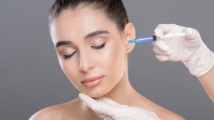
It was recommended that the patient be treated with a dermal filler to resolve both the tear trough deformity and the palpebromalar groove, but not the hyperpigmentation. For that, the patient was recommended to undergo further treatments. Administration was carried out with the patient sitting upright in her chair, with her eyes open throughout the procedure.
The filler of choice for this treatment was Teosyal Redensity II from the Teosyal fillers line. It was selected due to its low hygroscopic behaviour that is due in part to its composition, which is cross-linked and non-crosslinked hyaluronic acid at a 15mg/g concentration. This formulation results in minimal swelling in this area, especially when compared to other fillers. The palpebromalar groove was treated first, with small bolus injections that started at 0.5cm lateral to the lateral canthus and continued down the groove. A 30-gauge needle was used to administer the filler deep into the internal aspect of the orbital septum for volume creation.
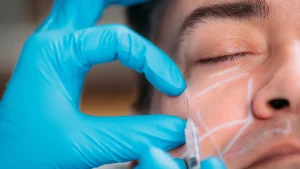
A needle is preferable to a cannula, as it is more precise. Approximately 0.1ml was used for each eye. After initial correction, it was noted that further correction of the palpebromalar groove was necessary above the orbicularis oculi muscle, but due to the risk of bruising, the remainder of the procedure was carried out with a cannula towards the end of the treatment process.
Upon treatment of the lateral aspect of the eye, the tear trough deformity was the next area of focus. Filler was placed deep into the suborbicularis oculi fat (SOOF) with a 25-gauge needle. This is typically not recommended; most practitioners advocate the supraperiosteal placement of filler in this area. However, as Teosyal Redensity II contains uniquely low hygroscopic properties, this filler should not produce overt swelling or puffiness.
Administration of filler was done using an intermittent retrograde micro-bolus technique that entailed the placement of the product along the length of the tear trough in positions level to the orbital rim. The entry point was 1cm from the end of the tear trough. After administering the product, the area was then gently massaged.
At this point in the procedure, all deep injections had been administered. As the patient had good skin thickness and elasticity, it was decided to inject more filler over the tear trough deformity and palpebromalar groove in the subcutaneous layer above the orbicularis oculi muscle. This was achieved using micro-fanning thread injections with the cannula. Provided the patient has adequate skin thickness, injecting the filler into the subcutaneous layer minimizes the risk of lumps forming post-injection. Continuous massage was employed during administration.
A total of 0.8ml of filler was used, with 0.4ml per eye. After the treatment, no ice packs or cooling were applied, as the patient showed no sign of post-operative bruising. The patient was advised to refrain from wearing makeup for the ensuing 24 hours and to avoid exercise and exposure to extreme temperatures for 48 hours post-treatment. A follow-up session was scheduled for 10 days after treatment, as the patient could not make the normal 14-day review. At the follow-up session, the patient reported that she was happy with the results. There was some residual mild swelling at the needle and cannula insertion sites. This was expected because in many cases it takes about three weeks for swelling to completely subside. The patient was seen again 14 months later for retreatment.
Conclusion
The under-eye area can be a difficult region to treat due to the associated high risk of complications, such as necrosis or blindness. Thus, any procedure involving this area should be performed by an advanced practitioner. Minimizing risk of complications can be achieved by undercorrection and a detailed knowledge of injection anatomy. Practitioners who are looking to perform these types of treatments should take steps to gain the necessary skill and knowledge, particularly in terms of injection techniques (especially those that are product-specific). The use of a cannula is absolutely necessary to attain positive outcomes when treating the tear trough area.